
Our efforts were focused on perfecting the performance of the imaging and high power probe for the sake of carrying out DQF-MT experiments on blood vessels. This probe was built in order to combine two features that are not found simultaneously in any commercial imaging probe. The first is a very homogenous RF field, that allows for the use of fast imaging methods such as RARE. These methods require the use of multiple pulses, hence the demand for perfect RF pulses. As mentioned in our application for the Slezak Fund, the performance of our probe in this regard is much more than satisfying.
The second feature for which the probe was built is to make the use of very short pulses, i.e. high-power pulses which are crucial for the DQF-MT experiments. The signal in the DQF-MT experiment is acquired from fast-relaxing macromolecules (Fig. 1), thus the excitation pulse for this magnetization must be short enough so as to minimize the signal loss due to relaxation during the pulse time.
Our first prototype of probe yielded a stable 90o pulse of 70ms. Further reduction of the pulse length by increasing the RF power resulted in a loss of capacitor due to breakdown voltage. In order to further reduce the pulse length we decided to utilize a transformer-like device that is based on a toroid. This transformer separates the circuit into a transmitting section, that carries the high voltage, and a tuning section, where tunable capacitors tune, match and balance the circuit via a reduced voltage. The price paid for this voltage reduction over the capacitors is the reduced tuning capability of these capacitors. However, this is not a problem, since our first prototype exhibited tuning capability far beyond the needs of any conceivable MRI experiment one would perform with this probe.
The choice of a toroid as a voltage divider was made due to its minimal radiation emission, hence its minimal distortion of the RF homogeneity and minimal loss of power.
As a result of the above modifications, the 90o pulse duration was reduced to 50ms. The extent of S/N improvement in the DQF-MT imaging method due to this reduction can be estimated from relaxation measurements of the double quantum signal of a blood vessel. The exponential signal decay constant found from fitting the curve at short decay times (fig 1), is 23ms. Thus, an estimate of 58% percent of a during-pulse S/N gain is achieved by shortening the pulse.
To sum up, a significant improvement in the expected image quality of blood vessels was achieved, without degrading any of the qualities of the probe.
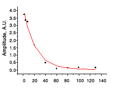 |
Fig. 1. Decay of the double quantum magnetization in a sheep carotid artery. The rate constant of decay measured for the first eight points was found to be 23ms. The 20ms reduction of pulse length, thus corresponds to an estimate of 58% of S/N gain in the DQF-MT signal during the pulse. |
