
Placental failure to supply oxygen and nutrients to the fetus and to remove toxic waste may result in a small for gestational age baby which is prone to higher risks of morbidity and mortality. Thus, knowledge of the governing parameters of feto-maternal blood circulation both in normal and pathological placentas is essential for developing reliable diagnostic and monitoring techniques. Power Doppler ultrasound measurements of the placental blood flow were performed in 20 low-risk pregnant women at 21-40 weeks of gestational age that were recruited for the research. The placental arteries were measured in the umbilical cord insertion into the placenta, at the first generation on the chorionic plate and at a depth of 1-1.5 cm inside the placenta by Sonoline Elegra ultrasound system (Siemens) and a multi-frequency probe (4.5-7.2 MHz). The blood flow velocity at peak systole (PS) and end diastole (ED), and Doppler indices (PI- pulsatility index, RI- resistance index, S/D-systole to diastole ratio) were measured. A regression line was drawn in all graphs in order to specify the trend of the results.
The results are presented from 20 to 40 weeks of gestation. The flow velocity at PS and ED increased towards term as shown in Figs. 1a and 1b, respectively. For all gestational age, the flow velocity was highest at the cord insertion, decreased in the chorionic blood vessels and was lowest in the intraplacental vessels. The velocity of blood flow in the chorionic vessels at 20 week of gestation was close to the velocity in the intraplacental vessels. As the week of gestation increases up to 40, the flow velocity increases towards that measured in the umbilical cord insertion. The Doppler indices S/D, PI and RI were highest at low gestational age, decreased as the gestational age decreased, and were the lowest at 40 week of gestation.
The placenta grows as the fetus develops. The blood vessels become wider, and more blood vessels are developed in order to perfuse the placenta. More blood is needed for the fetus is supported by the observations that the flow velocity at systole and diastole increased with the gestational age. The measured DIs of the various placental blood vessels showed similar trend as in the DIs tables used in obstetrics for the flow in the umbilical cord. The measurements of the flow velocity at PS and ED yielded better sensitivity than the DIs parameters since they are measured directly rather than presenting relations between the flow velocities and distributed along a longer range.
The pattern of the flow velocity at PS and ED, S/D and RI of the chorionic blood vessels are very interesting (Fig. 1, broken line). It may be that in early gestational age vessels in the placenta are not fully developed, and the flow is similar to those in the intraplacental blood vessels. Thus, obliterations of intraplacental vessels may be detected by measurements of the flow in the vessels of the chorionic plate. In late gestation, the blood flow, the placenta and the diameter of the umbilical cord increase. As a consequence, it may be that the diameter of the first generation in the intraplacental vessels increase in order to supply blood to the rest of the placenta.
 |
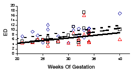 |
| Fig. 1: Flow velocities at PS (a) and ED (b): (i) umbilical cord in the insertion (diamonds), regression line dots, (ii) vessels in the chorionic plate (squares), regression line broken line, (iii) intraplacental vessels (triangles), regression line solid line. | |
