The evolvement of cardiac dysfunction in hypertensive heart disease and following myocardial infarction (MI) depends greatly on the degree of cardiac fibrosis. Production of type I collagen, the main component of the fibrotic lesion, is regulated by the renin-angiotensin-aldosterone system and blockade of the angiotensin II and aldosterone receptors has been shown to reduce collagen deposition in the heart. A crucial step in collagen deposition is the extracellular processing of it's precursor, procollagen, by procollagen C-proteinase (PCP), a reaction stimulated by the procollagen C-proteinase enhancer (PCPE) protein. We have previously shown that, in cultured rat heart fibroblasts, PCPE and collagen expression is enhanced by both aldosterone and angiotensin II. In the current study, we went on to examine whether aldosterone and angiotensin II are involved in the regulation of PCPE expression in vivo during ventricular remodeling post MI. Wistar rats were operated to create acute MI and allowed to recover with or without administration of the respective aldosterone and angiotensin II receptor antagonists, spironolactone (Spi, 25 mg/kg/d) and irbesartan (Irb, 80 mg/kg/d). Sham operated rats served as the controls. Five weeks post infarction the rats were sacrificed, their hearts were removed rapidly, and the expression of PCPE and collagen mRNAs in the surviving myocardium was assessed by Northern blotting and in situ hybridization. While the scars in the hearts of Irb-treated rats appeared similar in size to those observed in the untreated infarcted rats, the scars in Spi-treated animals were found to be considerably smaller. In situ hybridization demonstrated for the first time the presence of PCPE transcripts in the heart, which were localized to fibroblasts adjacent to the infarct scar (Fig. A). Collagen mRNA was found in the same region but the distribution of collagen-presenting cells was much wider, possibly because it's high abundance facilitated detection. The levels of PCPE and collagen mRNAs were reduced in the myocardium of Spi-treated rats, indicating that aldosterone coordinately regulates the expression of both proteins (Fig. B). In light of the well established role of angiotensin II in cardiac fibrosis, the lack of inhibition by irbesartan in this study was difficult to explain. Different treatment regimens may address this issue.
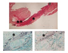
Fig. A. Upper panel, collagen staining by sirius red, *scar; ** fibrosis. Lower panel, fibroblasts containing collagen (left) and PCPE (right) mRNA; *scar region. |
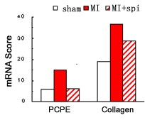
Fig. B. Bar graph summarizing the expression scores of PCPE and collagen.
|

