All patients are at risk for rejection, and must be carefully followed regardless of demographic variables. The surveillance for allograft rejection generally centers on the routine use of endomyocardial biopsies, accompanied by periodic physical examinations and echocardiography. We proposed to develop a new approach of examining tissue and to overcome the drawbacks of the standard biopsy diagnostic procedures used today. This approach is based on specifically binding of fluorescence marker on the lymphocytes and monocytes\macrophages. A few problems exist when trying to diagnose rejection through fluorescence biopsy. Specificity of binding, window of time for making the measurements and the mathematical reconstruction of the location of fluorophores. We have created a mouse model where we injected Ab-conjugated fluoropohres to the suspected area. We followed the washout of the fluorophores in order to distinguish between control and diseased mice. The results are shown in the graph of Fig. 1. Fluoresce image of a specifically bound fluorophores is shown in Fig. 2. We were able to reconstruct from images captured from excited fluorophores deeply embedded in tissue. These results enable us to continue to work on this method in order to finally achieve a minimal invasively method, of without the need of biopsy, to locate concentrations of lymphocytes and monocytes\macrophages which are present in the rejection area of an allograft.
Fig. 1. Washout experiments. |
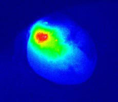
Fig. 2. Fluorescence image. |

