Impedance cardiography is the study of cardiac function determined from measurements of the electrical impedance of the thorax. Variations in tissue resistivity associated with physiological events, such as cardiac and respiratory activities, allows bio-impedance technique to be used as a diagnostic and monitoring tool. In the present study, a 2-dimensional model of the torso and a numerical method were used to investigate the changes in the potential distribution associated with physiological events, such as cardiac and respiratory activity. Transverse CT images of the torso were scanned. Noise reduction and contour following algorithms were applied to differentiate between different compartments in the torso. The extracted tissue types were lungs, blood, ribs, bone marrow of the cord, breast fat, skin, skeletal muscle, and heart muscle (Fig. 1). Isotropic homogeneous conductivity was assigned to each one of these compartments. The volume conductor problem was solved numerically using the finite volume method to determine the potential distribution developed due to dipole source (Fig. 2). Cases with different heart and lung volumes were examined to investigate the sensitivity of impedance techniques to detect cardiac output. Significant changes (Fig. 3) were detected in the potential distribution inside the volume conductor as a result of cardiac and respiratory activities indicating that the impedance technique employed in the present study has a very good promise in detecting cardiac output.

Fig. 1. Torso anatomy after segmentation, used as an input to the numerical model
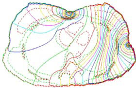
Fig. 2. Surface potential Vs. heart area, in different stages of the cardiac cycle. |
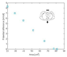
Fig. 3. Surface potential Vs. heart area, in different stages of the cardiac cycle. |

|
