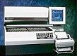
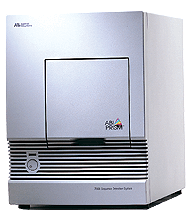
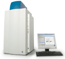
LS 6500
LS6500 Liquid Scintillation Counter
LS 6500 Scintillation Counter combine advanced scintillation
technology, configurability to meet your exact requirements and built-in
upgradability to add features, as you need them. Value Systems are instrument
configurations that meet standard requirements, H-Number Plus, our patented
quench quench monitor, and Versa-RackT are standard features on both LS 6500
Value Systems. You can count samples in any combination; up to 336 standard
vials (20 mL).
The isotope library C 14, H3, I125, .S35 and P32 are permanently stored
in the system.
Biolistic PDA
Gene Transfer
The biolistic PDS-1000/He unit. Top, main chamber
containing the micro carrier launch assembly and the bombardment helium
pressure gauge. The central gauge (in the left side of the instrument) monitors
the vacuum within the chamber, and the 2 lower knobs adjust the vacuum flow and
vent rates. The Helium metering valve is next to the main chamber.
PDS-1000 | He System

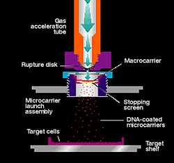
Transform Any Cell Type
The biolistic PDS-1000/He instrument is capable of accelerating sub
cellular-sized tungsten or gold microprojectiles coated with DNA over a range
of velocities and target distances necessary to optimally transform many
different cell types. Particle delivery with the PDS-1000/He system is a
convenient method for transforming intact cells in culture since very little
pre- or post bombardment manipulation is necessary, and sample containment
within the evacuated bombardment chamber offers maximum control.Biolistic
bombardment allows transformation of animal cells with unique growth
requirements that are not amenable to gene transfer using any other method.
Reports describing how to transform cultured mammalian cells with the
PDS-1000/He device (see References) show improved transient transformation of a
variety of sensitive mammalian cells that are poorly transformed by chemicals,
lipofection, or even electroporation. In addition, you can use less DNA and
fewer cells for either transient or stable transformation with the biolistic
particle delivery system. This technique is much easier and faster than the
tedious task of microinjecting embryos. How the Unit WorksThe helium pressure
and vacuum circuits in the PDS-1000/He system effectively accelerate the
microcarriers into the target cells: After all the materials are in place, the
chamber door is closed and a vacuum is applied. The vacuum reduces the
frictional drag on the DNA-coated microcarriers and provides a safety
interlock; the instrument cannot be activated unless a vacuum is
drawn.Activating the fire switch allows helium to flow into the gas
acceleration tube at a rate regulated by the helium-metering valve and
monitored by the helium pressure gauge. The gas is held until the burst
pressure of the rupture disk is reached. This generates a helium shock wave
into the bombardment chamber.The shock wave hits the microcarrier launch
assembly and propels a plastic macrocarrier holding DNA-coated microcarriers
toward the target cells. A stopping screen placed between the macrocarrier
assembly and the cells retains the plastic disk, while allowing the coated
microprojectiles to pass through and transform the target cells.Bombardment
ParametersWe recommend these settings for bombardment of a variety of cell
types. Many factors affect bombardment efficiency, but most users will find it
sufficient to optimize the major variables individually, then test their
interactions on a limited scale.
Bombardment Parameters
|
Cell Type
|
Vacuum(inches Hg)
|
TargetDistance (cm)
|
Helium Pressure (psi)
|
Particle Size
|
Bacteria
|
29
|
6
|
1,100
|
M5 tungsten
|
Yeast
|
28
|
6
|
1,300
|
0.6 µm gold
|
Algae
|
29
|
6
|
1,300
|
0.6 µm gold
|
Plant
|
Embryos
|
28
|
6
|
1,300
|
1.0 µm gold
|
Callus or cell cultures
|
28
|
9
|
1,100
|
1.0 µm gold
|
Sub cellular organelles
|
28
|
6
|
1,300
|
0.6 µm gold
|
Animal
|
Tissue cultures
|
15
|
3
|
1,100
|
1.6 µm gold
|
Tissue sections
|
25
|
9
|
1,100
|
1.6 µm gold
|
We recommend these settings for bombardment of a variety of cell
types. Many factors affect bombardment efficiency, but most users will find it
sufficient to optimize the major variables individually, then test their
interactions on a limited scale.
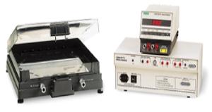
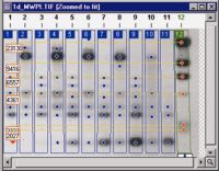
Chef Electrophoresis
The CHEF-DR II system effectively resolves DNA fragments in the
range of 5 kb to 6 Mb. This system lets you enhance resolution by entering two
blocks of running conditions to be executed successively. The CHEF-DR II system
exclusively employs the most common angle of electrophoresis for PFGE, 120°.
This system is simple to program, and has proven to be a solid performer.
Not Just Mapping Anymore
-
Strain Typing-Molecular Epidemiology
-
Apoptosis Assays
-
DNA Damages and Repair Studies
-
Large Protein Separations
-
Disease Loci Mapping
-
YAC, BAC, PAC, and Cosmid Mapping
-
Chromosome Rearrangements
-
PFLP and DNA Fingerprinting.
Confocal Microscop
LSM 510
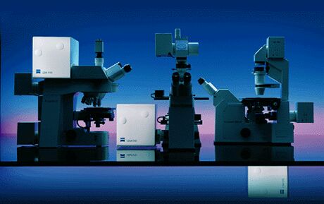
LSM 510 system is
unmatched. It allows multi fluorescence images to be collected without
compromising resolution and efficiency.
Imagine the new system for research:
Unprecedented flexibility for individual applications. Entirely new acquisition
and processing functions that guarantee fast and efficient structural and
functional analysis of your specimens. A stable and reliable system, allowing
you to concentrate on the content of your research
The ZEISS LSM system is highly integrated scanning module and fully motorized
microscope
Argon Laser: 458nm , 488nm and 514nm
HeNe: 543nm and 633nm
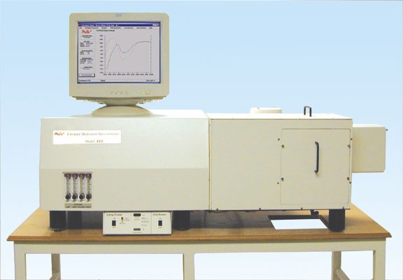
Circular Dichroism
Spectrophotometer
The Aviv Model 215
Circular Dichroism Spectrometer
Circular dichroism spectroscopy measures physical phenomena wherein
molecules absorb to different extents circularly polarized light. Differences
in the absorbance are directly related to the optical activity of chiral
molecules.
Circular dichroism is an essential tool of measuring secondary structure of
proteins, ligand binding and macromolecular association. Deconvolution of
the spectra can give a quantitive measure of protein secondary structure.
The Aviv model 215 Circular Dichroism Spectrometer records circular dichroism
as a function of wavelength, time, temperature, pH and concentration using a
double monochromator containing two UV grade prisms as dispersing elements.
Focusing optics allows use of micro cells. A 50 Khz photoelastic modulator
produces right and left circularly polarized light.
The instrument allows secondary structure estimates, stopped-flow kinetic
studies, wavelength scans, temperature scans, titration studies, total
fluorescence measurements, and more. Wavelength ranges 165nm to 900nm
.
Multiphor
MultiphorT II flatbed
Two-dimensional (2-D) electrophoresis is a powerful analytical
technique capable of resolving thousands of individual proteins in complex
biological samples. In cases where the resolution provided by larger-format
systems is not required, the speed and convenience of a mini-format can be
exploited.
The Multiphor T II Flatbed Electrophoresis Unit allows both the first and
second dimensions of 2-D electrophoresis to be performed with the same
instrument. Three second-dimension separations are run simultaneously on a
single second-dimension gel, which is particularly useful for comparative
studies, as all three samples experience identical separation conditions.
Silver staining using the PlusOne T Silver Staining Kit, Protein; and the
Hoefer T Automated Gel Stainer provides exceptionally sensitive visualization
when applied to the
ExcelGel T precast gel used in this technique.
Multiphor II electrophoresis system is a versatile modular system of Pharmacia
for horizontal electrophoresis, isoelectric focusing 2-D electrophoresis and
electrophoretic transfer.
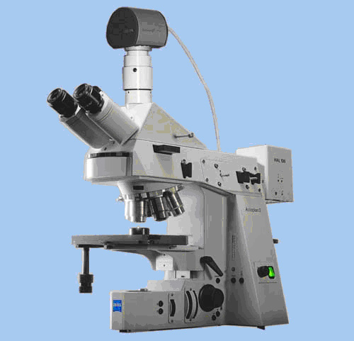
Axioplan
Zeiss Axioplan
Zeiss upright fluorescence microscope
The filters are:
1)UV filter, EX 365, EM 420.
2)Green filter, EX 490 EM 525.
3)Red Filter,EX 540 EM 580.
Fluorescence microscope
DMRBE
Leica DMRBE
Leica manufactured DMRB upright microscope with magnafire 12-bit
color CCD camera.
The filters are:
1)B.G.R filter for uv, blue and green, excitation filter, BP
400/20, Dichromatic mirror rkp 415, Suppression filter BP 465/20.
2)DAPI excitation filter BP 340-380, Dichromatic mirror rkp
400, Suppression filter LP 425.
3) M2(Rhodamin) excitation filter BP 546/14, Dichromatic
mirror rkp 580, Suppression filter LP 590.
4) Gfp excitation 470/40x, Dichromatic mirror
hq495LP, em hq 525/50m.
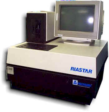
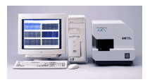
Gamma Counter
Packard
RIASTAR
Packard RIASTAR multi well gamma counter
5 well multi well gamma counter with multi channel analyzer technology The
instrument is configured with 10 detectors.
Packard RiaStar Gamma Counting Systems includes multi -channel analyzer,
Instrument Performance Assessment (a self-inspecting system) and automatic
protocol operating.
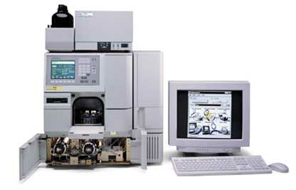
HPLC- High Performance Liquid
Chromatography
Waters HPLC
The water's hplc includes 50u pumps model 626, fluorescence detector
474, PDA detector 996, 600s controller, vacuum degasses and non-cooled auto
sampler 717.
The instrument is fully controlled by the millennium software.
Luminometer
TD-20/20 Luminometer
The Turner Designs model 20e is equipped with a Hamilton injector
for automatic injections.
Its EPROM includes auto ranging and 2nd functions for smoothing, pre delay,
delay, repeat, and continuos reading.
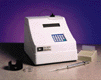
The most sensitive display range is 0.000 to 9.999 and the least sensitive
display range is 0000 to 9999.
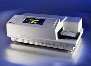
Microplate Reader
Spectramax 190
SPECTRAmax 190
The SPECTRAmax 190 spectrophotometer is ideal for most life science
applications, especially DNA analysis. For aqueous solutions, our patented*
PathCheck® sensor technology senses the depth of the liquid in each microplate
well, and normalizes the absorbance value to a 1-cm pathlength. The corrected
absorbance is within 5% of the value obtained in a conventional
spectrophotometer
The SPECTRAmax 190 microplate reader allows you to:
Use the extinction coefficient of your sample to calculate concentrations
directly,
Correct for volume differences in the wells, expand the dynamic range of your
assay,
Detect pipetting errors and check performance of your pipetting devices
Preparative Electrophoresis
Preparative
Electrophoresis
Any molecule that can be resolved in a slab gel can be purified
quickly, easily, and completely using continuous elution electrophoresis. Using
SDS-PAGE, native PAGE, or agarose gel electrophoresis, the Model 491 prep cell
and mini prep cell isolate individual components from crude or partially
purified samples. These instruments are ideal for use at any stage in a
purification scheme. As a final step following chromatography or preparative
isoelectric focusing, these instruments can be used to isolate a specific
molecule from any contaminants remaining in the sample.
· Conventional electrophoresis buffer systems and media are
used
· Purify nanograms to milligrams of a particular molecule
· Using SDS-PAGE, purify molecules that differ in molecular
weight by as little as 2%
· Using native PAGE, isolate molecules that differ in
isoelectric point by as little as 0.1 pH unit.
Purified molecules are automatically collected in individual liquid fractions
and are immediately available for sequence analysis, crystallography, antibody
production, enzyme kinetics studies, NMR, bioassays, or any other study where
homogeneous bio-molecules are required. Single- and double-stranded DNA and RNA
can also be purified using the prep cell.

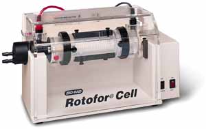
Rotofor
Protein Purification Simplified
The Rotofor system fractionates complex protein samples in free solution using
preparative isoelectric focusing. This nondenaturing technique rapidly isolates
proteins of interest from the bulk proteins in complex biological samples. With
the addition of the mini Rotofor focusing chamber, this unique system
accommodates a wide range of sample volumes and sizes.
-
Ideal for the isolation of uncharacterized proteins from crude or
partially purified samples
-
Interchangeable mini and standard focusing chambers
-
Process micrograms to grams of protein in 18 to 58 milliliters of
sample
-
Easy set-up and short run times enable 500-fold purification in less
than 3 hours
-
Ceramic cooling finger dissipates heat generated during high voltage
runs
-
The unique membrane core stabilizes proteins in 20 focused zones and
facilitates free-solution collection without mixing
Sequencer ABI 377 and 3100
http://www.tau.ac.il/lifesci/zabam/sequencing/
Telemeter
Ask Dr. Eli Geffen
Spectrophotometer
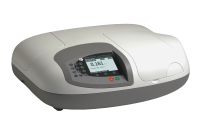
Ultrospec 2000
Pharmacies ultrospec 2000 UV/visible spectrophotometer measures
standard absorbance, concentration and transmittance and has stored parameters
for DNA, RNA and oligonucleotide quantification and purity checking as well as
for protein contamination measurements in nucleic acid solutions.
Kinetic is also possible with 6 cell changer.
Bandwidth of 3nm and stray light at 220nm < 0.012%T.
Ultracentrifuge -
OptimaT LE
The Beckman optima le70 is equipped with the following rotors: SW
28, SW41, SW50.1, SW60 and 70, 70.1Ti.
The max rpm are 70,000 and max vacuum is less than 20 microns
Elutriation Centrifuge -Avanti®
J-25
The Avanti series is a centrifuge that allow the centrifugal
elutriation of living cells
Cells are subjected to two forces within the separation chamber: the
centrifugal field and the viscous drag of the fluid following in the opposite
(centripetal) direction. Each cell tends to migrate to a zone where its
sedimentation rates are balanced by the flow rate of the fluid through the
separation chamber.
By increasing the flow rate of the elutriation fluid in steps or decreasing the
rotor speed, successive populations of relatively homogenous cell sizes can be
washed from the chamber.
Densitometer-image master
ImageMaster 1D Image Analysis Software
Image analysis is an integral part of most of today's Life Science
applications and at APB we have the most comprehensive range of software
products available. Under the Image Master label, we cater for all your
analysis needs, from the basic documentation of a routine purity screen to the
querying of entire projects of 2D gels for discovering protein expression
changes.
This site covers all your analysis needs for applications which include band
and spot quantitation, the study of complex band pattern matching in 1D gels,
examination of lane relationships between many gels, high throughput screening
techniques aimed at 2D proteomics projects and rapid comparison of macro
arrays.
Combined with the large range of image capture devices we offer for all types
of detection methods, we can assemble a complete system for exactly the
application with which you work, whether it be DNA or proteins, arrays, 1D or
2D gels, labeled or stained.
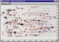
Umax scanner and two software for densitometry developed by Pharmacia enabling
quantitation of 1D gels and 2D gels
PAM 2000
"PAM-Fluorometer Family" employs the so-called
Pulse-Amplitude-Modulation (PAM) measuring principle, which is unique in
providing a highly selective measure of the relative chlorophyll fluorescence
quantum yield. In conjunction with the so-called "saturation pulse method" of
fluorescence quenching analysis
PAM-2000 -Fluorometers allow rapid assessment of
photosynthetic energy conversion. Measuring principle and quenching analysis
The Portable Chlorophyll Fluorometer PAM-2000 has been developed
on the basis of the well-known PAM Fluorometer. The patented pulse amplitude
modulation (PAM) measuring principle is used to measure the fluorescence yield
with high
Sensitivity and selectivity, whereby even strong unmodulated light (e. g. full
sunlight and saturation pulses with 10000 µmol quanta ms) causes no disturbing
effects.
The intensity of the measuring light is sufficiently low that it has no effect
on the Photosynthesis State and the fluorescence yield. In addition to the
current fluorescence yield (Ft, in continuous light) and the maximum yield
(Fm', in the saturation pulse) it is also possible to determine the minimum
yield (Fo', in the quasi-dark state). This information is automatically
evaluated by the data acquisition system integrated in the PAM-2000 Fluorometer
such that the quenching coefficients (qP and qN), the effective quantum yield
(F/Fm') and the apparent electron transport rate (PAR x F/Fm') are obtained
within seconds following each saturation pulse
IPGphor- Isoelectric Focusing System
IPGphor Isoelectric Focusing System is a dedicated instrument for
performing immobilized pH gradient (IPG) IEF. IPGphor delivers rapid,
high-resolution first dimension protein separations with pre-cast Immobiline
Dry Strip IPG gels.
The integrated system includes a programmable 8,000 V, 1.5 mA power supply and
Peltier solid-state temperature control (18-25°C ± 1 °C) for up to 12 IPG
strips.
IPGphor features an innovative strip holder, made out of aluminum oxide and
platinum, which allows reswelling of IPG strips and subsequent IEF separation
without handling the strips between steps. For examples of results obtained
using IPGphor Isoelectric Focusing System.
qRTPCR
The ABI Prism® 7000 Sequence Detection System is a complete,
real-time PCR system that detects and quantitates nulceic acid sequences. In
real-time PCR, cycle-by-cycle detection of accumulated PCR product is made
possible by combining thermal cycling, fluorescence detection, and
application-specific software in a single instrument. Quantitative results are
available immediately after PCR without additional purification or analysis.
Real-time, quantitative PCR applications include gene expression and
pathogen detection.Post-PCR detection is also available for non-quantitative
assays such as allelic discrimination (SNP detection) and plus/minus assays.
Key Features and Benefits
-
Multicolor detection provides flexibility for multiplex quantiation
assays, allelic discrimination assays, and plus/minus assays utilizing an
internal positive control (IPC)
-
Precision optics, combined with a sophisticated multicomponenting
algorithm, provide accurate, highly-reproducible results.
-
Small footprint facilitates easy placement in any laboratory
(notebook computer may be placed on top of the 7000 system when space is very
limited)
-
Peltier-based, 96-well block thermal cycling system is easy to use
with standard 96-well plates or 0.2 mL tubes
-
Proven assay development guidelines save time and money
Scanner 428
Affymetrix 428 Array Scanner
The Affymetrix 428 Array slides Scanner is a digital
confocal laser scanning epifluorescent microscope for viewing fluorescent-dye
tagged samples.
The scanner is equipped with two lasers : 532nm and a 635nm
(cy3 and cy5).
Data Mining Tool (DMT).
The Data Mining Tool (DMT) software provides a variety of tools
for filtering and sorting GeneChip® array data enabling you to find your most
significant data results quickly and easily. Key features include:
Pairwise statistical analysis |
| . |
Clustering |
| . |
Annotation
Integration
DMT is compatible with Windows NT and Windows 2000 operating systems.
FTIR
Nexus 470
Fourier
transform-infrared (FT-IR)
Fourier
transform infrared (FT-IR) is a technique that uses an interferometer for data
collection and a digital Fourier transformation to process the data.
TheNexus
FT-IR spectrometer family
provides superior performance to meet the demanding infrared sampling
requirements. Design excellence and smart system components ensure the
Nexus
will provide reproducible and accurate results. The
Nexus
provides real-time feedback on all aspects of sample analysis with exclusive
Enhanced Synchronization Protocol (E.S.P.) technology.
The
Nexus is Thermo Nicolet's research-grade, fully upgradeable FT-IR spectrometer.
The Nexus is the highest-performance spectrometer with superior design,
ease of use and total flexibility.
The
Nexus takes full advantage of Thermo Nicolet's unique Smart System technology
that features automatic purge, optical component recognition and much
more, providing unsurpassed accessibility to research level results! Every
Nexus system links all aspects of the system together so that experiments are
optimized. A Nexus spectrometer automatically configures sampling parameters,
beam path, spectral range and experiment types. Snap a Smart Accessory into
your Nexus and you will find uncompromising sampling performance. Thermo
Nicolet Smart Accessories provide the highest optical throughput,
reliability and sampling flexibility. OMNIC software provides the
interface that makes the Nexus smart system work. Our wide range of add-on
experiments, coupled with expanded spectral range, give spectroscopists options
available with no other instrument.
All
Nexus spectrometers can be configured for multiple spectral ranges. Changing
spectral range is as easy as removing an optical
component and placing the new one into its precision, pinned position.
Nexus automatically updates ranges, sensitivity and parameters. Thermo Nicolet
is the only FT-IR manufacturer with the proven ability to upgrade from a basic
system to a fully advanced research system. The universal optics and
electronics platforms make Nexus spectrometers flexible in every sense of the
word. Nexus components, including optics, are pinned-in-place, which ensures
they are always in alignment as well as guaranteeing reproducibility, stability
and the ability to swap and replace components.
The Nexus spectrometer has the most efficient optical path
of any infrared spectrometer, providing excellent quality spectral data. Nexus
systems utilize continuous dynamic alignment to ensure exceptional
high-resolution lineshapes, and provide superior
FT-IR spectrometers record the interaction of IR radiation with
experimental samples, measuring the frequencies at which the sample absorbs the
radiation and the intensities of the absorptions. This is a single beam
instrument.
Detector-DTGS (KBr Window): deuterated triglycine sulfate. A
very sensitive detector for mid infrared range measurement. Most routine
scanning instruments feature the more expensive and more sensitive DTGS
detector, which is one or two orders of magnitude more sensitive than lithium
tantalate, depending upon implementation. One drawback of the DTGS detector in
a process environment is that it stops functioning as a detector at
temperatures higher than 41 degrees C; the detector depolarizes above this
temperature.
Mid ***** 3.00 - 6.00 microns (3000-6000 nanometer),
Beamsplitter***KBr Spectral Range(cm-1)= 7400-350

PIT PIT tag (Passive
Integrated Transponder) externally mounted or implanted RFID (Radio Frequency
Identification) product used to positively identify animals and other objects
(rocks, golf balls, driftwood, etc.) for a lifetime. Each tag is a read-only
tag that is programmed to transmit a unique code only when activated. This code
cannot be changed. There are tags that can be programmed but Biomark, Inc. does
not offer that product nor do we recommend it for animal identification .
PIXImus
href="http://www.gemedicalsystems.com/rad/bonedens/peripheral/piximus_spec.html">PIXImus
Academic and pharmaceutical researchers commonly use the mouse for
investigations of genetics, cellular physiology, and of agents affecting bone
or soft-tissue composition. Bone densitometry and body composition measurement
using dual-energy x-ray absorptiometry (DEXA), eliminate the need for
destructive chemical analysis - a time- and labor-consuming process that
requires days or weeks to complete. Bone densitometry allows the researcher the
opportunity to make multiple measurements in-situ during the life of the
animal. Unfortunately, DEXA densitometers using rectilinear scanning require as
much as 30 minutes for a total body acquisition with relatively poor precision
(>5%) and spatial resolution (>0.5mm). The long process requires careful
sedation techniques and often endangers animal safety. The PIXImus densitometer
provides bone mineral and body composition results from total body imaging in
under 5 minutes. Fast imaging allows use of milder sedation that is safer on
animals.The PIXImus allows accurate and precise measurement of bone and tissue
for small animals 10-50 g (e.g.,mice, lemmings). This measured bone exhibits
excellent correlation to total ashed weight (r=0.99). PIXImus uses a lower
x-ray energy than that used for peripheral densitometry in humans in order to
achieve contrast in the extremely low density bone. Excellent precision of BMD
and %Fat makes PIXImus ideal for longitudinal studies. The standard total body
focuses on the active bone area in the subcranial region. There are manual
ROI's for selected regions within the total body image, such as spine and
femur. /P>< /FONT>
Microplate Luminometer

Features
Detection limit of 3 x 10-21 moles of luciferase using
Promega's Luciferase Assay Reagent
-
Linear dynamic range greater than nine decades
-
Crosstalk better than 3 x 10⁻⁵
-
Dual injector
-
Microplate 96-well format
-
Easy to use software
Synergy

When making fluorescence determinations, the Synergy HT uses a tungsten halogen
lamp with interference filters:
Default Excitation Filters No.1: POS1=340/11,
POS2=485/20, POS3=530/25 and POS4=
310/20.
Optional Excitation Filters
No.2 : POS1= 360/40, POS2= Plug , POS3=Plug and POS4=
540/35.
Default Emission Filters No1:
POS1=460/40, POS2= 528/20, POS3=590/20
and POS4= 580/50.
Optional Emission Filters No2:
POS1=Empty, POS2= 645/15 , POS3= 400/10 and POS4=600/40 .
When absorbance measurements are made, the instrument switches to a Xenon
Flash Lamp and a monochromator for wavelength selection from 200 to 999 nm.
Features
-
Automatic top and bottom epifluorescence read modes optimized for both
sensitive homogeneous and cell-based assays
-
Software-controlled automatic probe height adjustment with built-in plate
sensor to prevent probe crashes
-
Scanning monochromator for wavelength scans to identify peak absorbance and
provide full spectral analysis
-
Luminescence measurements with top detection for best sensitivity
-
Reads 6- to 384-well plate formats including PCR trays and Terasaki plates
-
Well area scanning for popular cell-based assays
-
Time Resolved Fluorescence capability
-
USB and serial ports for easy connectivity
-
Includes powerful PC software GEN 5 with exclusive PowerReports feature for
custom reporting
VP-ITC
VP-ITC

Features include:
-
Fixed-in-place cells for reproducible ultrasensitive performance
with low maintenance
-
Active cell volume ~ 1.3 ml
-
Cells fabricated from Hastelloy®, which has excellent chemical
resistance properties
-
Linear drive liquid delivery system incorporating a precision 250
microliter syringe
-
Variable mixing speed
-
Temperature range 2ºC to 80ºC
-
Internal Peltier mechanism - no external heating or cooling required
-
Directly measure binding constants in the range of <
102 to ~109 M
-1
-
Measures tight binding constants (greater than 109 M-1)
using competitive binding techniques
-
NEW! Now
includes Single Injection Method software.
X-ray Diffraction

The possibility of using
powder diffraction methods to study materials was recognized shortly after the
discovery of X-ray diffraction by von Laüe and Knipping in 1910. In particular,
the construction of a simple powder diffractometer was described by Hull in
1917, and the instrument was used to obtain patterns from a number of simple
materials such as diamond, graphite and iron. Within a few years, many others,
including the Braggs and Pauling, had exploited the powder method to study a
wide range of materials, including metals, minerals, and simple organic solids
.
X-ray Diffraction System for
Protein Crystals
X-Ray1
1. Rigaku (manufacturer); ultraX18 (18kW rotating anode x-ray
generator); Raxis-IV Image Plate area detector;
confocal "blue" OSMIC X-ray mirrors.

X-Ray2-NEW

2 . Rigaku (manufacturer); MicroMaxTM-007 HF
(800W rotating anode microfocus x-ray generator);
R-AXIS IV++
Image Plate area detector; VariMaxTM focusing X-ray mirrors.
LSM_META
The Scanning module of LSM 510 META
| Details |
|
|
| The unique scanning module is the core of the LSM 510 META.
It contains motorized collimators, scanning mirrors, individually adjustable
and positionable pinholes, and highly sensitive detectors including the META
detector. All these components are arranged to ensure optimum specimen
illumination and efficient collection of reflected or emitted light. A highly
efficient optical grating provides an innovative way of separating the
fluorescence emissions in the META detector. The grating projects the entire
fluorescence spectrum onto the 32 channels of the META detector. Thus, the
spectral signature is acquired for each pixel of the scanned image and
subsequently can be used for the digital separation into component dyes. |
The Beam Path:

1.
Optical Fibers |
2.
Motorized collimators |
3. Beam combiner |
4.
Main dichroic beamsplitter |
5.
Scanning mirrors |
6.
Scanning lens |
7.
Objective lens |
|
8. |
Specimen |
9.
Secondary dichroic beamsplitters |
10.
Confocal pinhole |
11.
Emission filters |
12.
Photomultiplier |
13.
META detector |
14.
Neutral density filters |
|
15. |
Monitor diode |
16.
Fiber out
ND1000
The NanoDrop ND-1000 UV-Vis Spectrophotometer enables highly accurate
analyses of 1 ul samples with remarkable reproducibility. The patented sample
retention system eliminates the need for cuvettes and capillaries which
decreases the measurement cycle time. In addition, the high absorbance
capability eliminates the need for most dilutions. You'll find yourself making
fewer dilutions and taking more readings than ever before, essentially becoming
a more quantitative laboratory.
The ND-1000 offers many benefits: Small samples: designed for 1 ul samples
Large dynamic range: measures 2-3700 ng/ul< (dsDNA) on a single sample. No
need to change out cuvettes or caps. High absorbance capability: 50 times that
of traditional spectrophotometers No Dilutions for most samples No extra costs
for consumables Full Spectrum (220-750nm) 10 second measurement time No
cuvettes or capillaries Sample is recoverable Small size (size of a tissue box)
Easy set up (plug and play) Software designed for life sciences Free software
upgrades
FL3-11
FL3-11
 Modular Spectrofluorimeter: single grating excitation and emission
Monochromators, Datamax/32 software with GRAMS license .
Modular Spectrofluorimeter: single grating excitation and emission
Monochromators, Datamax/32 software with GRAMS license .
SPECIFICATIONS:
OPTICS: All reflective for focusing at all wavelengths and precise
imaging for micro samples
SOURCE: Ozone-free 45O W xenon lamp eliminates venting
SPECTROMETERS: Plane grating Czerny-Turner design maintains focus at all
wavelengths
EXCITATION: Range 200-950 nm, optimized in the UV, blazed angle 330 nm
EMISSION: Range 200-950 nm, optimized in the visible, blazed angle 500nm
BANDPASS: Ex.& Em, 0-30 nm, continuously adjustable
WAVELENGHT ACCURACY : +/- 0.5nm
STEP SIZE: 0.0625 -100 nm
SCAN SPEED: 150 nm/s
INTEGRATION TIME: From 1 ms to 160s
EMISSION DETECTOR: Photomultiplier range 200-850 nm
REFERENCE DETECTOR: Photodiode selected for stability
SAMPLE MODULE: T-box design to allow second detection emission channel
WATER RAMAN SIGNAL: min. 400.000cps, EX=350nm, EM=397nm, BP=5 nm,
integration=1s
S/N RATIO: 4000/1 minimum Thermocouple Drive Single Position-Includes
controller, software, stirring mechanism and nitrogen cord for flushing: Range:
-10 to Required serial port in the PC. Polarization Accessory L-format, fully
automated dual polarizer. Located at both the entrance and exit of the T-box
for optimum extinction ratio and Sensitivity. Sample, Compartment, Accessory,
Electronics Necessary to control and manipulate automated sample chamber
accessories.
FLA 5100
FLA 5100 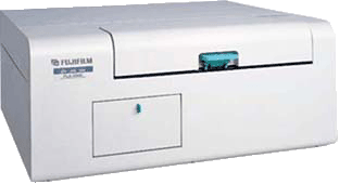
Radioisotopic Imaging
The system ensures radioisotopic imaging with high sensitivity, which comes from
Fujifilm's unique and reliable technologies. Imaging plate (IP) consists of a
radiosensitive layer of photostimulable phosphor crystals on a polyester
support. The IP accumulates irradiated radiation energies on exposure for
further scanning excitation process. The innovation and ergonomic design
provides a broad range of advantages.
Fluorescence
FLA-5100 has three kinds of excitation laser beams: 473nm, 532nm and
635nm. The system has four filters (510nm, 575nm,
665nm, and a filter for IP) for upgrading flexibility.
MALS
| DAWN® HELEOST II
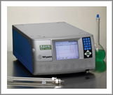
18-angle light scattering detector for the measurement of absolute molecular
weight, size, and conformation of macromolecules in solution. The DAWN®
HELEOST II may be used in batch mode (off-line) or connected on-line to an
HPLC/HPSEC/AFFF, etc.. The DAWN® HELEOST II utilizes a 120mW
solid-state laser operating at 658 nm and also has on-board digital signal
processing hardware for up to four external devices such as RI and/or UV
detectors. Also contains an analog output from the 90° detector for interfacing
to strip chart recorders. The DAWN® HELEOST II has a gorgeous 64,000
color LCD display, thermostatic control options, depolarization options, and
the ability to interface to high temperature GPC systems like the PL210 and
Waters Alliance 2000. On-board analog-to-digital conversion
with Ethernet communication for data acquisition
Optilab T-rEX
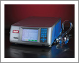
Optilab Refractive Index Detector
The Optilab® rEX (refractometer with EXtended range) is an Refractive Index
detector with 256 times the detection power and up to 50 times the
dynamic range of every RI detector in existence today. Using a
combination of cutting-edge semiconductor photodiode technology and proprietary
computer algorithms, the rEX presents a host of instrument firsts.
For example, there are no range or gain settings. The full range of
instrument detection is always present, and the full sensitivity exists over
the entire range.
The rEX also has a flow cell with a total volume of only 7.4 uL -- 25% less
than the leading refractometers available today. This translates into minimized
band broadening and better temperature stability.
The Optilab® rEX can also do something no other on-line Refractive Index
detector can: it can measure the absolute refractive index of a solution. In
addition, it can measure the dn/dc of a solvent at the same wavelength
of light as the light scattering instrument. And just to make sure it can't be
matched, you can replace the light source in the rEX for other wavelengths
(e.g. 690, 633, 488, etc.)!
The new Optilab® rEX shares the ability its predecessor (the Optilab® DSP) to
go to below ambient temperatures as easily as above ambient. No hassle
temperature control can be programmed down to 4°C or as high as 50°C.
Compatible with all HPLC systems.
FACS
BD Biosciences
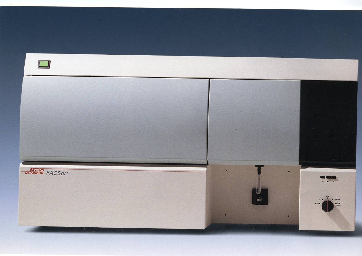
Two Lasers: 488 and 630
Software:
Flow Jow
ChirascanT
Circular Dichroism Spectrometer

Specifications
| Light source
|
150W
Xe arc, air-cooled
150W Hg-Xe arc, air-cooled (optional)
|
| Monochromator
|
F/7 double
prism, both dispersive, both polarising
|
| Wavelength
accuracy
|
±0.2 nm (170nm - 400nm), ±0.5 nm (400nm -
900nm)
|
| Wavelength
precision
|
±0.05 nm (170 - 400 nm), ±0.1 nm (400 - 800
nm)
|
| Wavelength
resolution
|
0.1nm
throughout range.
|
| Bandwidth |
0 to 2nm at 160nm, 0 to 4nm at 180nm, 0 to
>10nm at >250nm
|
| Wavelength
range
|
165nm - 1360nm.
Detector option for >900nm
|
| Stray
light
|
<5ppm at 200nm, <8ppm at 180nm
|
| Baseline
stability
|
±0.02 m o /hr
|
| Scanning
modes
|
Discrete sampling step-scan, Adaptive sampling
step-scan
|
| Scanning
speeds
|
Dependent upon
step-scan resolution approximately 500nm min-1 at 0.5nm step
|
| Kinetic
modes
|
Linear timebase, Split-linear timebases,
Logarithmic timebase
|
| Response
time
|
Not
applicable. Chirascan uses the superior method of digital oversampling which
eliminates the risk of damage to data.
|
| CD full scale
|
±1500 m
|
| CD resolution
|
64 bit
|
| RMS
noise
|
0.015m ° at 185nm, 0.012m ° at 200nm, 0.015m °
at 500nm, (1nm bandwidth, 16s oversampling (integration))
0.007m ° at 185nm, 0.007m ° at 200nm, 0.008m ° at 500nm, (2nm bandwidth, 16s
oversampling (integration))
|
| Data
channels
|
Internal: 2 standard; up to 16 optional.
External (standard): 6 inputs for
temperature (multiplexed); 3 analogue inputs; plenty of digital I/O.
|
OptimaT L-90 K Preparative Ultracentrifuge

The L-90 K instrument iscapable of generating
694,000 x g at speeds up to 90,000 rpm, the Optima L-90 K
Ultracentrifuge enables you to perform more separations in less time. This
versatile floor model operates with a broad range of superb rotors, including
zonal and continuous flow for large-volume separations.
The L-90 K offers the reliability of a
vacuum-encased induction drive, the simplicity of user-friendly,
microprocessor-based control, and environmentally-friendly cooling systems that
eliminate the need for CFCs and other harmful liquid refrigerants. All
this makes the L-90 K the right choice for your laboratory's routine separation
needs.
OptimaT TLX Personal Benchtop Ultracentrifuge
The Optima TLX features maximum performance capabilities of
657,000 x g and sample volumes ranging from 0.2 mL to 5.1 mL per tube.
System features include imbalance-tolerant, direct
vacuum-encased, air-cooled drives, rotor imbalance detection, moisture-purging
vacuum pump,solid-state temperature control systems; automatic rotor
identification, user diagnostics; programmability with up to five step changes,
ten selectable acceleration/deceleration rates and microprocessor control..

Specifications
|
|
Optima TLX (627,000 x g; 120,000 rpm) |
| Speed control |
± 50 rpm of set speed |
| Sample imbalance tolerance |
Up to 10% of volume in opposite tubes |
| Drive cooling* |
Air-cooled* |
| Refrigeration system* |
Solid state, no CFCs/ODCs* |
| Set temperature range |
2°C to 40°C in 1°C increments |
| Temperature control |
± 2°C of set |
| Ambient operating range |
15°C to 35°C |
| User-settable programs |
10 user programs; all user programs
have up to five steps |
| Acceleration/Deceleration rates |
10 Accel/11 Decel |
| Moisture-purging vacuum system |
Yes |
| Heat output |
0.6 kW/hr (1,750 BTU/hr) |
| Sound level 3 ft (1 m) away |
< 60 dB |
| Remote control via RS-232 |
Optional |
| HEPA filter |
Optional |
| Dimensions H x W x D in (mm) |
21 x 29 x 23 (533 x 737 x 584) |
| Weight |
178 lb (81 kg) |
Rotors:
-
TLA-120.2 Rotor, Fixed Angle,
Titanium, 10 x 2.0 mL, 120,000 rpm, 627,000 x g.
-
TLA-120.1 Rotor Package, Fixed Angle, Titanium, 14 x 0.5
mL, 120,000 rpm, 627,000 x g.
-
TLA-100 Rotor, Fixed Angle, Titanium, 20 x 0.2 mL, 100,000 rpm,
436,000 x g.
-
TLS-55 Rotor, Swinging Bucket, 4 x 2.2 mL,
55,000 rpm, 259,000 x g.
ImageQuant 350
ImageQuant 350 allows high-resolution scans of monochromatic
fluorescence gels, Coomassie Blue-stained gels, and other colorimetric
detection methods.
ImageQuantT 350 is a high-performance 16-bit cooled CCD camera-based system
designed for a wide range of monochromatic imaging applications. ImageQuant 350
is well-suited for quantitative analysis of Western blots. The system is also
fully capable of imaging monochromatic fluorescent gels and membranes,
CoomassieT Blue-stained gels, and other colorimetric detection methods.
Cultures and microplate-based assays can also be imaged. Whereas film is
valuable and convenient for certain applications, ImageQuant 350 supports a
more quantitative approach for a broader range of applications without
compromising sensitivity, resolution, or simplicity of operation. Most commands
are performed by pushing a single button, thereby supporting many different
applications in a day.
The ImageQuant 350 is a compact, easily installed system, designed for easy
exchange of cameras, lenses, and filters. The standard system includes one
manual fixed lens and one manual zoom lens, and the motorized system includes
one motorized fixed lens and one manual zoom lens.
The standard UV transilluminator and the white light source (a fold-down table),
as well as any optional light sources, are housed dust-free inside the cabinet.
The UV transilluminator can be operated at either 302 nm or 365 nm at
low or high intensity. For cutting sample bands from the gel, the
transilluminator slides out of the cabinet.
Using ImageQuant TL analysis software with ImageQuant 350 provides high levels
of automation, accuracy and reduces analysis time to a minimum.
AUC
Analytical ultracentrifuge
Analytical ultracentrifugation is used for the determination of
molecules masses in solution. The instrument analyzes macromolecular
sedimentation over a wide range of conditions such as
concentration,temperature, ionic strength and pH.
Sedimentation velocity and
sedimentation equilibrium experiments can be done with the
instrument. The operating methodology measures the
relative change in the distribution of molecular weights, providing an
efficient way to measure heterogeneity, stoichiometry and self-associating
systems. The XL-A/XL-I's measurements are based on the first principles of
thermodynamics and hydrodynamics, no standards or calibrations are
required.
|
|
All data and images were taken
from the companies sites.
This page
was last updated on:

;)
)